
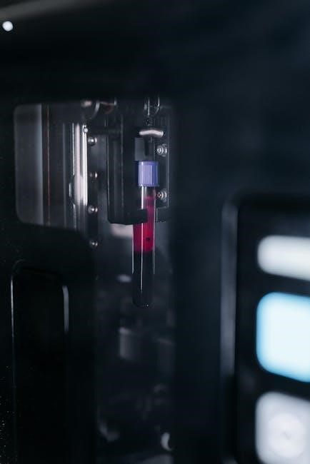
Blood collection tubes, distinguished by a color-coding system, are vital for sample integrity. Each tube’s cap indicates its specific additives and intended clinical tests, ensuring accurate diagnoses.
Importance of Proper Tube Selection
Correct blood tube selection is paramount for reliable lab results. Utilizing the wrong tube can compromise sample integrity, leading to inaccurate diagnoses and potentially incorrect treatment plans. Each tube contains specific anticoagulants or additives designed for particular tests – from hematology to coagulation studies.
The color-coded system isn’t merely for organization; it dictates the tube’s functionality. Failing to adhere to this system introduces variables that invalidate test outcomes, impacting patient care significantly. Proper selection guarantees sample stability during collection, transport, and analysis.
Color-Coding System Overview
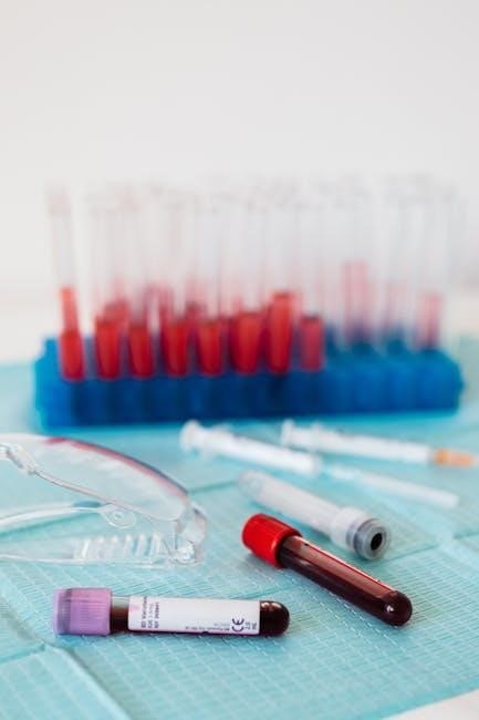
The blood collection tube color-coding system is a standardized method for identifying tube contents. Each color denotes specific anticoagulants, additives, and appropriate test types. This visual cue minimizes errors and ensures compatibility between the sample and the required analysis.
Understanding this system is crucial for phlebotomists and healthcare professionals. Tubes range from light blue for coagulation studies to lavender for hematology, each designed to preserve sample integrity and deliver accurate results. Consistent adherence to this system is vital.
Common Blood Collection Tube Colors and Their Uses
Blood vacutainer tubes vary in color, each designed for specific tests. These color-coded tubes contain different additives to stabilize samples for accurate clinical diagnosis.
Light Blue Top Tubes
Light blue top tubes contain sodium citrate, functioning as an anticoagulant. This prevents blood clotting, making them essential for coagulation studies, like prothrombin time (PT) and activated partial thromboplastin time (PTT). The citrate binds calcium, halting the coagulation cascade. Proper filling is crucial for accurate results; an incorrect blood-to-additive ratio can skew testing outcomes. These tubes are vital for assessing a patient’s clotting ability and monitoring anticoagulant therapy.
Uses for Coagulation Studies
Light blue top tubes are specifically designed for coagulation assessments, including tests like PT, PTT, and fibrinogen levels. These evaluations are critical for diagnosing bleeding disorders and monitoring the effectiveness of anticoagulant medications such as warfarin or heparin. Accurate results depend on precise blood-to-additive ratios and proper handling. These studies help clinicians understand a patient’s ability to form blood clots and manage related risks effectively.
Sodium Citrate as an Anticoagulant
Sodium citrate, within the light blue top tubes, functions as an anticoagulant by binding calcium, which is essential for the coagulation cascade. This prevents clotting, allowing for accurate assessment of coagulation factors. The ratio of citrate to blood is crucial; too little yields inaccurate results, while too much prolongs clotting times. It’s vital to avoid contamination during collection to maintain sample integrity for reliable testing.
Red Top Tubes
Red top tubes, lacking any additives, are incredibly versatile for serum collection and a broad range of general chemistry tests. Allowing the blood to clot naturally before centrifugation yields serum, free from cellular components. This makes them suitable for tests like electrolytes, glucose, and lipid panels. Their adaptability makes them a staple in many clinical settings, offering broad diagnostic capabilities.
Serum Collection and General Chemistry Tests
Red top tubes facilitate serum collection, crucial for numerous general chemistry tests. After clotting, centrifugation separates serum from cells, enabling accurate analysis. Common tests include measuring electrolytes, glucose, cholesterol, and kidney/liver function markers. The absence of additives prevents interference, ensuring reliable results. This simplicity and broad applicability make red top tubes a cornerstone of routine clinical diagnostics.
No Additives – Versatility in Testing
Red top tubes, lacking any anticoagulants or additives, offer exceptional versatility in laboratory testing. This absence minimizes interference with various assays, making them suitable for a broad spectrum of clinical chemistry analyses. They are ideal when specific additive requirements aren’t mandated, simplifying the testing process and reducing potential complications. Their adaptability makes them a fundamental tool in many diagnostic settings.
Lavender/Purple Top Tubes
Lavender or purple top tubes contain EDTA (ethylenediaminetetraacetic acid), a potent anticoagulant crucial for hematology applications. EDTA prevents blood clotting by binding calcium, preserving cell morphology for accurate blood counts and differential analyses. These tubes are commonly utilized for CBCs, blood smears, and immunohematology tests. Proper mixing – approximately eight inversions – is essential for effective anticoagulation.
EDTA Anticoagulant – Hematology Applications
EDTA functions as a powerful anticoagulant by chelating calcium ions, effectively halting the coagulation cascade. This makes purple top tubes ideal for hematology tests where maintaining blood in a liquid state is critical. Applications include complete blood counts (CBCs), blood smears for morphological evaluation, and hemoglobin A1c assays. EDTA preserves cellular components, ensuring reliable and accurate results in hematological analyses.
Common Tests Performed with Purple Top Tubes
Purple top tubes, containing EDTA, are frequently utilized for a broad spectrum of hematological assays. These include routine complete blood counts (CBCs), assessing red blood cell, white blood cell, and platelet levels. Furthermore, they support blood smears for microscopic examination and hemoglobin electrophoresis. Proper mixing – typically eight inversions – is crucial for effective EDTA distribution and reliable test outcomes.
Gray Top Tubes
Gray top tubes contain potassium oxalate and sodium fluoride, uniquely designed for glucose testing. Potassium oxalate binds calcium, preventing coagulation, while sodium fluoride inhibits glycolysis – the breakdown of glucose by enzymes. This preservation is critical, especially when samples cannot be processed immediately, or travel long distances. Utilizing gray top tubes ensures accurate glucose measurements, avoiding falsely lowered results.
Potassium Oxalate and Sodium Fluoride – Glucose Testing
Gray top tubes utilize a specific combination: potassium oxalate halts clotting by removing calcium, and sodium fluoride acts as an antiglycolytic agent. This prevents glucose consumption by blood cells, ensuring accurate results, particularly when analysis is delayed. These tubes are ideal when samples aren’t immediately processed, maintaining glucose integrity during transport and storage for reliable testing.
Preserving Glucose Levels for Accurate Results
Gray top tubes are specifically designed for glucose testing, preventing its decline post-collection. The sodium fluoride within inhibits glycolysis – the breakdown of glucose by cells. This is crucial when samples cannot be analyzed immediately, or when drawn distantly from the lab. Using gold SST or red top tubes can lead to inaccurate readings due to glucose metabolism.
Green Top Tubes
Green top tubes contain heparin, an anticoagulant preventing blood clotting, making them ideal for emergency chemistry and blood gas analysis. Heparin doesn’t interfere with most clinical tests, supporting diverse applications. These tubes are vital for specialized testing requiring whole blood, offering a reliable sample for accurate and timely diagnostic results in critical care settings.
Heparin Anticoagulant – Emergency Chemistry and Blood Gas Analysis
Heparin’s primary role is preventing blood coagulation, crucial for emergency chemistry panels and precise blood gas analysis. It doesn’t affect most tests, offering broad compatibility. This makes green top tubes essential for stat testing, providing rapid results for critical patient care decisions. Heparin’s effectiveness ensures sample integrity for accurate assessments.
Applications in Specialized Testing
Heparin tubes extend beyond basic analysis, supporting specialized tests like plasma volume determination and certain immunological assays. Their anticoagulant properties preserve sample components for detailed evaluations. They are also utilized in situations requiring whole blood analysis, maintaining cellular integrity. This versatility makes green-top tubes invaluable in diverse laboratory settings, aiding complex diagnostic procedures.
Specialized Blood Collection Tubes
Specialized tubes, like gold SST and black top, offer unique features for specific tests. These enhance serum quality or facilitate precise ESR measurements, improving diagnostic accuracy.
Gold/SST Top Tubes
Gold or Serum Separator Tubes (SST) are widely utilized for various clinical chemistry tests. These tubes contain a gel separator that spins out during centrifugation, creating a clear separation between the serum and cellular components. This enhances serum quality, minimizing interference and improving the accuracy of test results. They are versatile, suitable for a broad range of analytes, and are a common choice in most clinical laboratories due to their reliability and ease of use.
Serum Separator Tubes – Enhanced Serum Quality
Serum Separator Tubes (SSTs) utilize a gel barrier to efficiently separate serum from cells after centrifugation. This physical barrier prevents prolonged contact between the serum and cellular components, minimizing the release of substances that could interfere with testing. The resulting high-quality serum exhibits reduced hemolysis and clotting factors, leading to more accurate and reliable analytical results in various clinical chemistry assays.
Applications in Various Clinical Chemistry Tests
Gold/SST tubes are extensively used in a broad spectrum of clinical chemistry tests. These include lipid profiles, cardiac markers, liver function tests, kidney function assessments, and comprehensive metabolic panels. The superior serum quality obtained from these tubes ensures accurate measurements of analytes, aiding in the diagnosis and monitoring of diverse medical conditions. Their versatility makes them a cornerstone of modern laboratory diagnostics.
Black Top Tubes
Black top tubes contain sodium citrate, primarily utilized for ESR (Erythrocyte Sedimentation Rate) testing. This test measures how quickly red blood cells settle in a tube over time, indicating inflammation within the body. Proper filling is crucial for accurate results. These tubes prevent blood clotting, allowing for reliable assessment of erythrocyte sedimentation, a valuable diagnostic tool.
Sodium Citrate – ESR Testing
Sodium citrate, within black top tubes, prevents blood coagulation by binding calcium, essential for the clotting process. This allows for accurate ESR (Erythrocyte Sedimentation Rate) measurement. The ESR test detects inflammation, aiding in diagnosing conditions like arthritis or infections. Precise blood-to-anticoagulant ratios are vital for reliable results, ensuring accurate clinical assessments.
Determining Erythrocyte Sedimentation Rate
The Erythrocyte Sedimentation Rate (ESR) measures how quickly red blood cells settle in a tube over one hour. Elevated rates indicate inflammation within the body, prompting further investigation. Sodium citrate tubes are crucial, preventing clotting and ensuring accurate sedimentation. This non-specific test aids in diagnosing conditions like infections, autoimmune diseases, and certain cancers, guiding clinical decisions.
Pink Top Tubes
Pink top tubes contain EDTA, making them essential for immunohematology applications, specifically blood grouping and compatibility testing. These tests are vital before blood transfusions to prevent adverse reactions. EDTA preserves cell morphology, ensuring accurate antigen identification. Proper collection and handling are critical for reliable results, safeguarding patient safety during transfusion procedures.
EDTA – Blood Grouping and Compatibility Testing
EDTA in pink top tubes prevents clot formation, preserving blood cell integrity for accurate blood grouping. This is crucial for determining a patient’s blood type (A, B, AB, or O) and Rh factor. Compatibility testing then ensures donor blood won’t trigger an immune response. Precise antigen identification relies on EDTA’s morphological preservation, guaranteeing safe blood transfusions.
Immunohematology Applications
Pink top tubes containing EDTA are foundational in immunohematology, the study of blood groups and transfusion medicine. They facilitate antibody screening and identification, vital for preventing transfusion reactions. Accurate red blood cell phenotyping, determining specific antigen profiles, relies on preserved cell morphology. These tests are essential for managing patients with complex transfusion needs and autoimmune hemolytic anemias.
Understanding Anticoagulants and Additives
Anticoagulants like EDTA, sodium citrate, and heparin prevent clotting, while sodium fluoride inhibits glycolysis, preserving sample integrity for accurate test results.
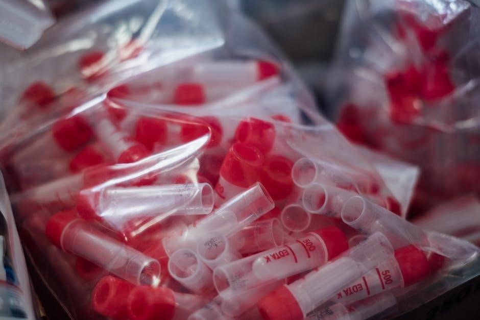
EDTA (Ethylenediaminetetraacetic Acid)
EDTA is a powerful anticoagulant binding calcium ions, halting the coagulation cascade. Commonly found in lavender/purple-top tubes, it’s ideal for hematology and molecular diagnostics. Proper mixing – around eight inversions – is crucial for effective anticoagulation. EDTA’s blood-binding capacity allows radiolabeling for renal function scans. It’s utilized for complete blood counts, ensuring reliable cellular analysis and preventing sample degradation during transport and storage.
Mechanism of Action and Common Uses
EDTA functions by chelating calcium ions, essential for coagulation factors. This prevents clot formation, preserving blood cells in their native state. Purple-top tubes utilizing EDTA are widely used for CBCs, blood typing, and hematological analyses. Its ability to preserve cell morphology makes it suitable for investigations requiring accurate cell counts and identification. Furthermore, EDTA’s versatility extends to immunohematology and viral marker studies.
Sodium Citrate
Sodium citrate acts as an anticoagulant by binding calcium, preventing the coagulation cascade. Light blue-top tubes containing sodium citrate are specifically designed for coagulation studies, like PT and PTT. This ensures accurate assessment of clotting factors. It’s also utilized in ESR testing with black-top tubes, allowing for reliable measurement of erythrocyte sedimentation rate, crucial in detecting inflammation.
Role in Coagulation Testing
Sodium citrate’s primary function is to prevent blood clotting during collection, making it ideal for coagulation tests. By chelating calcium ions – essential for the coagulation cascade – it effectively halts the process. Light blue-top tubes utilizing sodium citrate ensure accurate PT, PTT, and fibrinogen assessments. Proper fill volume is critical for maintaining the correct anticoagulant-to-blood ratio, guaranteeing reliable results.
Heparin
Heparin, a potent anticoagulant, is frequently used in green-top tubes for blood gas analysis and emergency chemistry panels. It inhibits thrombin, preventing clot formation without altering sample constituents significantly. This makes it suitable for tests requiring plasma, like arterial blood gases and electrolyte measurements. However, heparin can interfere with certain assays, so careful consideration is needed.
Applications in Blood Gas Analysis
Heparinized tubes, specifically those with a green top, are essential for accurate blood gas analysis. They prevent clotting, preserving the blood’s pH and gas tensions (oxygen and carbon dioxide). This is critical for assessing respiratory function and metabolic status. Samples must be analyzed promptly to minimize alterations, providing vital data for critical care and emergency medicine.
Sodium Fluoride
Sodium fluoride, found in gray top tubes alongside potassium oxalate, acts as an antiglycolytic agent. It inhibits glycolysis, the process where glucose is metabolized by blood cells, ensuring accurate glucose measurements. This is particularly important when samples aren’t immediately analyzed or are transported over longer distances, preventing falsely low glucose results.
Inhibition of Glycolysis in Glucose Testing
Sodium fluoride’s primary role is to inhibit glycolysis, the metabolic pathway breaking down glucose. Without it, glucose levels would decrease as blood cells consume it post-collection. Gray top tubes, containing sodium fluoride, preserve glucose integrity, delivering reliable results, especially when analysis is delayed. This ensures accurate diagnoses, particularly in glucose tolerance tests and routine screenings.
Blood Collection Best Practices
Proper technique – including order of draw, mixing, and storage – is crucial. These steps minimize contamination and maintain sample integrity for reliable results.
Order of Draw
Following the correct order of draw is paramount to prevent cross-contamination of additives between tubes, which can significantly skew test results. Generally, tubes with no additives are drawn first, followed by tubes with anticoagulants, and finally, tubes with additives like oxalate-fluoride. This sequence minimizes interference and ensures the accuracy of various hematological, coagulation, and chemistry analyses. Adhering to established guidelines, like those from CLSI, is essential for reliable diagnostic data.
Minimizing Contamination and Ensuring Accurate Results
Contamination from improper technique or incorrect order of draw can drastically impact test outcomes. Thoroughly clean the venipuncture site and avoid carrying over additives from one tube to the next. Gentle mixing immediately after collection distributes the anticoagulant evenly, preventing clotting. Proper handling and storage maintain sample integrity, leading to reliable and accurate laboratory results crucial for patient care and diagnosis.
Proper Mixing and Inversion
Adequate mixing is paramount post-collection to ensure the anticoagulant distributes evenly throughout the blood sample. Typically, gentle inversion – around 8-10 times – is recommended, especially for lavender/purple top tubes containing EDTA. Avoid vigorous shaking, which can cause hemolysis. Proper mixing prevents clot formation and guarantees accurate test results, particularly in hematology and coagulation studies, directly impacting diagnostic reliability.
Ensuring Adequate Anticoagulant Distribution
Complete mixing guarantees the anticoagulant effectively prevents clotting. Insufficient distribution leads to micro-clot formation, compromising sample integrity and skewing results, especially in hematology tests. Gentle, consistent inversions – typically 8-10 times – are crucial. This process activates the anticoagulant’s mechanism, like EDTA’s calcium binding, ensuring reliable data for accurate clinical assessments and diagnoses.
Storage and Transportation
Maintaining sample integrity during storage and transport is paramount. Temperature control is vital; follow laboratory guidelines for each tube type. Prompt delivery to the lab minimizes analyte degradation. Proper labeling with patient details and collection time is essential. Protect tubes from physical damage and exposure to direct sunlight, ensuring reliable test results and accurate patient care.
Maintaining Sample Integrity
Sample integrity hinges on correct tube selection, proper mixing, and adherence to the order of draw. Avoiding contamination is crucial; gentle inversion ensures adequate anticoagulant distribution. Timely analysis or appropriate storage prevents analyte degradation. Following these steps guarantees reliable and accurate laboratory results, directly impacting patient diagnosis and treatment plans.
Pediatric and Low-Volume Tubes
Pediatric tubes, available in 2mL or less volumes, address smaller patient needs. These specialized tubes ensure accurate testing with minimal blood draws.
Availability of Smaller Volume Tubes
Recognizing the unique needs of pediatric and neonatal patients, manufacturers now widely offer blood collection tubes in reduced volumes. These typically range from 2mL down to even smaller sizes, accommodating the limited blood volumes obtainable from these vulnerable populations. This availability ensures that essential diagnostic testing can be performed without excessive blood loss, minimizing patient discomfort and potential complications. The smaller tubes maintain the same color-coding and additive principles as standard-sized tubes, ensuring compatibility with existing laboratory protocols and accurate test results.
Considerations for Pediatric Blood Collection
Pediatric blood collection demands extra care and technique. Utilizing smaller volume tubes is crucial to avoid iatrogenic anemia. Gentle venipuncture, appropriate site selection, and minimizing tourniquet time are essential. Distraction techniques can reduce anxiety. Always adhere to established protocols and institutional guidelines for pediatric phlebotomy, prioritizing patient comfort and safety. Proper tube selection, based on the required tests, remains paramount, mirroring adult practices but scaled to the child’s physiological capacity.